Our Vision: Innovating Bioburden Micoroscopy


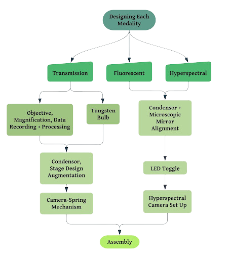
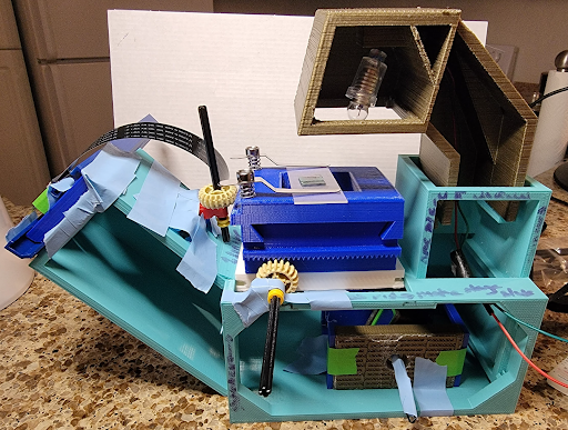
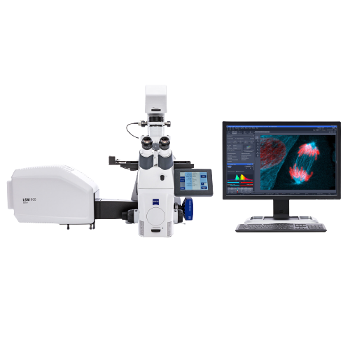
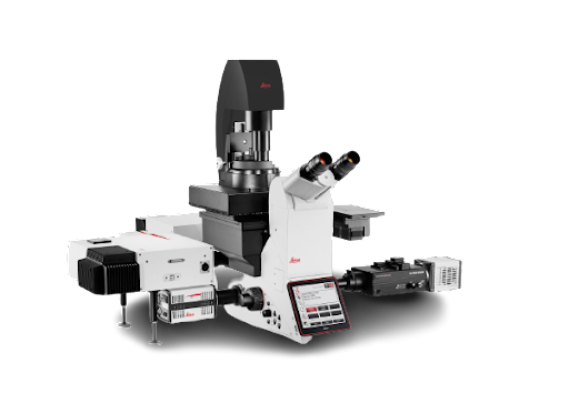
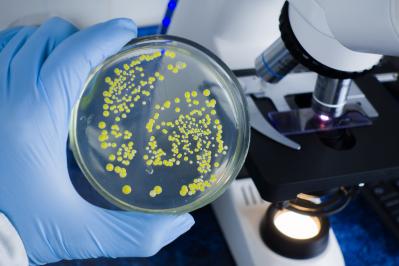
Every year, the Centers for Disease Control and Prevention reports that one in six Americans fall ill to foodborne illnesses. [1] The effects of foodborne illnesses can span from mild discomfort to serious, life-threatening consequences. Currently, diagnostic tools for pathogens require extensive travel time between the site of collection and observation, disrupting the quality and timeliness of analysis. Moreover, the equipment used has a limited range of analysis and adds up to billions of dollars in economic loss each year. We aim to reduce the frequency of these cases with our trimodal microscope and broaden the impact of analysis capabilities in microbial contamination testing. Our work builds upon current single or bimodal microscopes that have limited range of image analysis capabilities. Our novelty is light source shareability between the fluorescent and hyperspectral modes in arrangement with typical microscopy components housed in 3D printed casing. We have developed a transmission microscope designed to detect fluorescence in hyperspectral mode. The design consists of transmission illumination in an upright configuration, while the hyperspectral and fluorescence illumination are designed for forward detection. The two light pathways pass through a housing component that will allow users to easily switch between the modes. This novel, low-cost configuration will allow users to image samples for microbial contamination on-site for common pathogens such as E.coli and Salmonella, and receive feedback on the contamination levels to minimize the presence of pathogens on food we eat.
Overall, our trimodal microscope helps culitvators and suppliers along the food supply chain who want to understand and identify diseased food production by reducing test processes complexity, expense, and increasing access to onsite analysis. [2]

Next

Our Device

Our Design

The above flowchart describes our process into developing our device from seperate parts into one functioning device. As we have two light sources, we have made a two branch design plan. The transmission branch is straightforward with us finding an objective and condenser combination that produces good quality images while minimizing the microscope’s height. For the fluorescence and hyperspectral branch, we plan to use LEDs as they offer a sufficient level of illumination for the sample. In terms of imaging, we aim to implement the use of a hyperspectral camera to output high quality images. Coupled with the imaging, we aim to implement a bluetooth system in order to allow images to be uploaded to a shared cloud. From the shared cloud, users would be able to share and access images gained from our microscope from anywhere at anytime.

Current Prototype

Shown above is our prototype. The outer casing is entirely made up of 3D printed parts that holds all the internal components together. It also has a stage that can move in the X and Y direction to allow the user to analyze the sample from different angles.

The transmission lightway for our device is very similar to a standard microscope capable of transmission imaging. It is set up to be a top-down orientation similar to to the diagram shown above. However our device places the light source in the top of the microscope as shown in the tungsten bulb in the photo of our prototype.

The other light pathway that our device will utilize is for the hyperspectral and flourescence imaging. This configuration is powered by an LED-pixel array [3] in combination with a light detector that is located on the right-most compartment of the device. The goal for this pathway is to output a continous spectrum of light in contrast with the segmented bands of light found in other types of imaging. This will be done through the LED-pixel array that allows for the output of multiple wavelengths of light. The last piece of the light pathway is found within the housing of the "guts". Within this housing, a gimble mechanism is found. With this gimble mechanism, the user would be able to shift the reflective mirrors that determine whether the transmission pathway would be used or the flourescent and hyperspectral pathway would be used. [4] This gimble mechanism would also prevent both modes from being used simultaneously.

Image Analysis

Cameras Used

In order to view and save images that can be used for further analysis, specific cameras can be used. Currently, there are two different options in order to fulfill this role: the Raspberry Pi camera and a 32 channel hyperspectral camera.

Next

Competitor Analysis

Zeiss LSM 980, Leica TCS SP8

Olympus IXPlore, Leica DMi8

Foldscope


Device Comparison

Device Name Modalities Cost
Foldscope Transmission $15-$50
Olympus Ixplore, Leica DMI8 Transmission and Flourescence $32,000
Zeiss LSM 980, Leica TCS SP8 Transmission, Fkourescence, and Hyperspectral $500,000 - $1,000,000
Our Device: Spectrinity Microscope Transmission, Flourescence, and Hyperspectral $1250

Our device offers many advantages There are three main competitors to our device. The first device is the Zeiss LSM 980, Leica TCS SP8. It is the most similar to our device in terms of integration as it includes all of transmission, hyperspectral, and flourescence modalities. In addition it uses a laser. However, it far more expensive than our device costing $500k to 1 million dollars. Our next competitor is the Olympus IXPlore, Leica DMi8. It is still expensive than our current device, at a cost of $32,000. However in comparison to our device with our three integrated modes, the Olympus IXPlore only offers two modes, being transmission and flourescence. Finally, our last main competitor is the Foldscope. Compared to our device, it is way more cost-effective, ranging from a price range of $15-$50. However it is very simplistic in its imaging analysis, as it only offers transmision capabilities. Overall, our spectrinity micorscope offers the best available device in terms of its low cost as well as its three integrated modes.

Next

Market

Target Market

We are currently developing our microscope for the bioburden market with our current interation of our device targeting the ever-growing agricultural food safety testing indsutry.

Market Size

Another reason why we chose the food safety testing market is because it is a fast-growing industry. In 2022, it had a worth of $21.1 billion dollars and has a projected worth of $31.1 billion dollars by 2027. It is a very lucrative and important market for us to target.

Specific Target Groups


Farms
Within the realm of the food safety industry, there are different stages where our microscope can be applicable. First at the farms specifically, there are the farms where the produce is first grown. If farmers were to detect any abnormalities or defects in their crops or seeds, they would be able to microscopically image these defects and upload them to the cloud or shared drive. Once uploaded, remote researchers would be able to evaluate and give farmers preventative actions if they are needed.

Government Agencies/Testing Sites
In terms of government agencies, the FDA conducts random crop sampling throughout the year in order to monitor crop health and growth. Similar to the farms, if the FDA workers were to find an anomaly or defect in the crops or seeds that they sample, they would be able to instantly get feedback from remote researchers through the cloud. A big advantage that this immediate feedback method through the cloud is that it would prevent sample contamination. Sample contamination can arise when a sample that is needed for analysis is contaminated from the source of the sample to the place for analysis. Contamination can alter the physical structure of the sample and even on the chemical level as well.

Wholesale Retailers
Finally, our microscope can be used to detect pathogens and other defects at factories. Once food is made at the factories, there are many modifications to the food that can be made such as manufacturing, processing, and packaging. Thus, there are many avenues in which food can be contaminated with pathogens or other unsafe and unwanted byproducts. With our device, workers would be able to prevent foods that are unhealty to be consumed by the public to be released to restaurants and grocery stores alike.

Next

Team Info

Hannah Ruth Kan
Team Lead, Software Designer

Jessica Lam
Creative Director

Isabella Jimenez
Prototype Developer

Alexander Lucas
Designer

Casey Park
Marketing Advisor


Check out Team Invismind's Linktree with the QR Code Above!

Next

References

  1. “Fast Facts About Food Poisoning.” Center for Disease Control and Prevention. https://www.cdc.gov/foodsafety/food-poisoning.html (accessed February 7, 2023)
  2. Recent advances of Hyperspectral Imaging Technology and Applications in agriculture.” Remote Sensing, 12(16), 2659. https://doi.org/10.3390/rs12162659
  3. Bosch, L. van den. (2020). Hyper- and multispectral imaging. Retrieved from https://wiki.tum.de/display/zfp/Hyper-+and+multispectral+imaging
  4. Tian, S., & Xu, H. (2022). Non Destructive Methods for the Quality Assessment of Fruits and Vegetables Considering Their Physical and Biological Variability. Food Engineering Reviews, 14,1-28. https://doi.org/10.1007/s12393-021-09300-0

Next